Multimedia Gallery
Lymphatics and the breast
The body is mostly composed of fluids. All its cells contain and are surrounded by fluids. In addition, four to five liters of blood circulate through the cardiovascular system at any given time. Some of that blood escapes from the system as it passes through tiny blood vessels called capillaries in the body tissues. Fortunately, there is a "secondary circulatory system" that reabsorbs escaped fluid and returns it to the veins.
That system is the lymphatic system. It runs parallel to the veins and empties into them. Lymph forms at the microscopic level. Small arteries, or arterioles, lead to capillaries, which in turn lead to small veins, or venules. Lymph capillaries lie close to the blood capillaries, but they are not actually connected. The arterioles deliver blood to the capillaries from the heart, and the venules take blood away from the capillaries. As blood flows through the capillaries it is under pressure. This is called hydrostatic pressure. This pressure forces some of the fluid in the blood out of the capillary into surrounding tissue. Oxygen from the red blood cells, and nutrients in the fluid then diffuse into the tissue.
Carbon dioxide and cellular waste products in the tissue diffuse back into the bloodstream. The capillaries reabsorb most of the fluid. The lymph capillaries absorb what fluid is left.
Edema, or swelling, occurs when fluid in or between the cells leaks into the body tissues. It is caused by events that increase the flow of fluid out of the bloodstream or prevent its return. Persistent edema may be a sign of serious health problems and should be checked by a health care professional.
The lymphatic system can play a very worrisome role in the spread of breast cancer.
Lymph nodes filter the lymph as it passes through the system. They are located at specific points throughout the body such as in the armpits and high in the throat.
Lymphatic circulation in breast tissue helps regulate the local fluid balance as well as filter out harmful substances. But the breast's lymphatic system can also spread diseases such as cancer through the body.
Lymphatic vessels provide a highway along which invasive cancerous cells move to other parts of the body.
The process is called metastasis. It can lead to the formation of a secondary cancer mass in another part of the body.
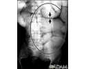
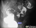
This mammogram shows a tumor and the lymph vessel network it has invaded.
No woman is too young to know that regular breast self-examinations can help to catch tumors earlier in their growth, hopefully before they spread or metastasize.
Lymphatics and the breast
Review Date: 7/23/2024
Reviewed By: Linda J. Vorvick, MD, Clinical Professor, Department of Family Medicine, UW Medicine, School of Medicine, University of Washington, Seattle, WA. Also reviewed by David C. Dugdale, MD, Medical Director, Brenda Conaway, Editorial Director, and the A.D.A.M. Editorial team.
© 1997- A.D.A.M., a business unit of Ebix, Inc. Any duplication or distribution of the information contained herein is strictly prohibited.
Animations
Illustrations
- 17 week ultrasound
- 30 week ultrasound
- Abdominal organs
- Abdominal quadrants
- Abdominal ultrasound
- Abdominal ultrasound
- Abnormal discharge from the...
- Actinic keratosis - close-up
- Actinic keratosis - ear
- Actinic keratosis on the arm
- Actinic keratosis on the fo...
- Actinic keratosis on the scalp
- Acute lymphocytic leukemia ...
- Acute monocytic leukemia - skin
- Adenocarcinoma
- Adenocarcinoma - chest x-ray
- Adrenal gland biopsy
- Adrenal gland hormone secretion
- Adrenal metastases - CT scan
- Adrenal Tumor - CT
- Aging spots
- Anatomical landmarks adult ...
- Antibodies
- Aortic aneurysm
- Aortic rupture - chest x-ray
- Appendicitis
- Ascites with ovarian cancer...
- Auer rods
- Barium enema
- Barium ingestion
- Basal cell cancer
- Basal Cell Carcinoma - close-up
- Basal cell carcinoma - close-up
- Basal Cell Carcinoma - face
- Basal cell carcinoma - nose
- Basal cell nevus syndrome
- Basal cell nevus syndrome -...
- Basal cell nevus syndrome - face
- Basal cell nevus syndrome -...
- Basal cell nevus syndrome -...
- Basophil (close-up)
- Before and after hematoma repair
- Benign juvenile melanoma
- Benign tumor of the skin
- Bile pathway
- Biopsy catheter
- Bladder biopsy
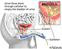
- Bladder catheterization - female
- Bladder catheterization - male
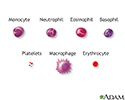
- Blood cells
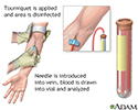
- Blood test
- Bone biopsy
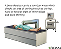
- Bone density scan
- Bone marrow aspiration
- Bone marrow from hip
- Bowen's disease on the hand
- BPH
- Brain
- Brain wave monitor
- Brain-thyroid link
- Breast lumps
- Breast pain
- Breast self-exam
- Breast self-exam
- Breast self-exam
- Bronchial cancer - chest x-ray
- Bronchial cancer - CT scan
- Bronchoscope
- Bronchoscopy
- Bronchoscopy
- Burns
- Calculating body frame size
- Candida - fluorescent stain
- Candidal esophagitis
- Canker sore (aphthous ulcer)
- Carotid duplex
- Carotid stenosis - X-ray of...
- Carotid stenosis - X-ray of...
- Carpal biopsy
- Carpal tunnel syndrome
- Causes of breast lumps
- Causes of breast lumps
- Central nervous system and ...
- Cervical biopsy
- Cervical cancer
- Cervical cancer
- Cervical cryosurgery
- Cervical cryosurgery
- Cervical neoplasia
- Cervical polyps
- Changes in face with age
- Changes in lung tissue with age
- Changes in skin with age
- Cheilitis - actinic
- Child thyroid anatomy
- Cholesterol producers
- Chromosomes and DNA
- Chronic lymphocytic leukemi...
- Chronic myelocytic leukemia
- Chronic myelocytic leukemia
- Chronic myelocytic leukemia...
- Cirrhosis of the liver
- Clubbing
- Coal worker's lungs - chest...
- Coal workers pneumoconiosis...
- Coal workers pneumoconiosis...
- Coal workers pneumoconiosis...
- Coal workers pneumoconiosis...
- Coccidioidomycosis - chest x-ray
- Cold cone biopsy
- Cold cone removal
- Colon culture
- Colonoscopy
- Colonoscopy
- Colposcopy-directed biopsy
- Congenital nevus on the abdomen
- Cryoglobulinemia of the fingers
- CSF cell count
- CSF protein test
- CT scan
- Cystography
- Cystoscopy
- Diet and disease prevention
- Digestive system
- Digestive system organs
- Donor liver attachment
- Emphysema
- Endocrine glands
- Endometrial biopsy
- Endometrial biopsy
- Endometrial cancer
- Enlarged spleen
- Epithelial cells
- ERCP
- ERCP
- Esophagus and stomach anatomy
- Ewing sarcoma - x-ray
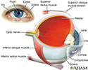
- Eye
- Facial drooping
- Fat tissue biopsy
- Fatty liver - CT scan
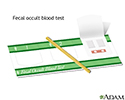
- Fecal occult blood test
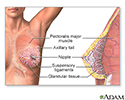
- Female breast
- Female breast biopsy
- Female perineal anatomy
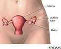
- Female reproductive anatomy
- Female reproductive anatomy
- Female urinary tract
- Fibroadenoma
- Fibrocystic breast change
- Fibroid tumors
- Formed elements of blood
- Gallbladder anatomy
- Gallbladder endoscopy
- Gallium injection
- Gallstones
- Gallstones, cholangiogram
- Gum biopsy
- Hairy cell leukemia - micro...
- Half and half nails
- Hand X-ray
- Head and neck glands
- Headache
- Heart - front view
- Heart - section through the...
- Heartburn prevention
- Hemangioma - angiogram
- Hemangioma - CT scan
- Hemoglobin
- Hemorrhoids
- Hepatocellular cancer - CT scan
- Hepatomegaly
- Hodgkin's disease - liver i...
- Hydrocele
- Hysterectomy
- Ileus - x-ray of bowel dist...
- Ileus - x-ray of distended ...
- Immune system structures
- Incision for lung biopsy
- Incision for pleural tissue...
- Incision for thyroid gland ...
- Intraductal papilloma
- Intravenous pyelogram
- Intussusception - x-ray
- Jaundice
- Jaundiced infant
- Kaposi sarcoma - close-up
- Kaposi sarcoma - lesion on ...
- Kaposi sarcoma - perianal
- Kaposi sarcoma on foot
- Kaposi sarcoma on the back
- Kaposi's sarcoma on the thigh
- Keratoacanthoma
- Keratoacanthoma
- Kidney - blood and urine flow
- Kidney anatomy
- Kidney function
- Kidney function tests
- Kidney metastases - CT scan
- Kidney tumor - CT scan
- Kidneys
- Koilonychia
- Large intestine (colon)
- Large intestine anatomy
- Lentigo - solar on the back
- Lentigo - solar with erythe...
- Leucine aminopeptidase urin...
- Lichen planus on the oral mucosa
- Lipoma - arm
- Liver biopsy
- Liver cirrhosis - CT scan
- Liver function tests
- Liver metastases, CT scan
- Liver with disproportional ...
- Lobes of the brain
- Lower digestive anatomy
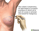
- Lumpectomy
- Lung biopsy
- Lung cancer - chemotherapy ...
- Lung cancer - frontal chest...
- Lung cancer - lateral chest...
- Lung mass, right lung - CT scan
- Lung mass, right upper lobe...
- Lung mass, right upper lung...
- Lung nodule - front view ch...
- Lung nodule, right lower lu...
- Lung nodule, right middle l...
- Lung tissue biopsy
- Lung with squamous cell can...
- Lungs
- Lymph node metastases, CT scan
- Lymphangiogram
- Lymphatic system
- Lymphoma, malignant - CT scan
- Malaria, microscopic view o...
- Malaria, photomicrograph of...
- Male reproductive anatomy
- Male reproductive system
- Male urinary system
- Male urinary tract
- Malignancy
- Malignant melanoma
- Malignant teratoma
- Mammary gland
- Mammogram
- Mammography
- Mediastinoscopy
- Megaloblastic anemia - view...
- Melanoma
- Melanoma - neck
- Melanoma of the liver - MRI scan
- MIBG injection
- Mongolian blue spots
- Mouth anatomy
- Mouth sores
- MRI scans
- Multiple basal cell cancer ...
- Muscle biopsy
- Nail infection - candidal
- Nasal biopsy
- Neck lump
- Needle biopsy of the breast
- Nerve biopsy
- Neuroblastoma in the liver ...
- Neurofibromatosis I - enlar...
- Non-small cell carcinoma
- Normal external abdomen
- Normal female breast anatomy
- Normal lung anatomy
- Normal lungs and alveoli
- Normal uterine anatomy (cut...
- Onycholysis
- Open biopsy of the breast
- Oral anatomy
- Oral thrush
- Oropharyngeal biopsy
- Oropharynx
- Osteogenic sarcoma - x-ray
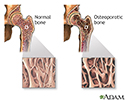
- Osteoporosis
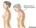
- Osteoporosis
- Osteoporosis
- Ovalocytosis
- Ovarian cancer
- Ovarian cancer dangers
- Ovarian cancer metastasis
- Ovarian cyst
- Ovarian cysts
- Ovarian growth worries
- Pancreas
- Pancreatic cancer, CT scan
- Pancreatic pseudocyst - CT scan
- Pancreatic, cystic adenoma ...
- Pap smear
- Pap smear
- Pap smears and cervical cancer
- Parathyroid biopsy
- Pelvic adhesions
- Pelvic laparoscopy
- Peritoneal and ovarian canc...
- Physical activity - prevent...
- Phytochemicals
- Pleural biopsy
- Pleural cavity
- Pleural smear
- Pleural space
- Primary and secondary hypot...
- Primary brain tumor
- Prostate cancer
- Prostate cancer
- Prostate gland
- PSA blood test
- Ptosis - drooping of the eyelid
- Pulmonary mass - side view ...
- Pulmonary nodule - front vi...
- Pulmonary nodule, solitary ...
- Quitting smoking
- Radiation therapy
- Rectal biopsy
- Rectal cancer - x-ray
- Red blood cells - elliptocytosis
- Red blood cells - multiple ...
- Red blood cells - normal
- Red blood cells - sickle an...
- Red blood cells - sickle cells
- Red blood cells - spherocytosis
- Red blood cells, sickle cell
- Red blood cells, target cells
- Red blood cells, tear-drop shape
- Renal biopsy
- Respiratory cilia
- Respiratory system
- Retina
- Salivary gland biopsy
- Sarcoid, stage II - chest x-ray
- Sarcoid, stage IV - chest x-ray
- Scrotal mass
- Secondhand smoke and lung cancer
- Selenium - antioxidant
- Sentinel node biopsy
- Serotonin uptake
- Sigmoid colon cancer - x-ray
- Sinuses
- Skeletal spine
- Skeleton
- Skeleton (posterior view)
- Skin
- Skin cancer - close-up of l...
- Skin cancer - close-up of l...
- Skin cancer - malignant melanoma
- Skin cancer - melanoma supe...
- Skin cancer - raised multi-...
- Skin cancer - squamous cell...
- Skin cancer, basal cell car...
- Skin cancer, basal cell car...
- Skin cancer, basal cell car...
- Skin cancer, basal cell car...
- Skin cancer, close-up of le...
- Skin cancer, melanoma - fla...
- Skin cancer, melanoma - rai...
- Skin cancer, melanoma on th...
- Skin cancer, squamous cell ...
- Skin layers
- Skull of an adult
- Small bowel obstruction - x-ray
- Small cell carcinoma
- Small intestine
- Small intestine biopsy
- Smoking hazards
- Smoking hazards
- Spermatocele
- Spinal tumor
- Spleen and liver metastases...
- Splenomegaly
- Squamous cell cancer
- Squamous cell carcinoma
- Squamous cell carcinoma - i...
- Stages of cancer
- Sternum - view of the outsi...
- Stomach
- Stomach cancer, x-ray
- Stomach ulcer, x-ray
- Structure of the colon
- Sun protection
- Sunburn
- Sunburn
- Sunburn
- Superficial anterior muscles
- Surface anatomy - normal palm
- Surface anatomy - normal wrist
- Swollen lymph nodes in the groin
- Swollen lymph nodes under arm
- Synovial biopsy
- Temperature measurement
- Teratoma - MRI scan
- Testicular anatomy
- Testicular biopsy
- Testicular ultrasound
- The large intestine
- Thermometer temperature
- Throat anatomy
- Thyroid cancer - CT scan
- Thyroid cancer - CT scan
- Thyroid enlargement - scintiscan
- Thyroid gland
- Thyroid gland biopsy
- Thyroid ultrasound
- Tobacco and cancer
- Tobacco and chemicals
- Tobacco and vascular disease
- Tobacco health risks
- Tongue
- Tongue biopsy
- Tooth anatomy
- Tuberculosis, advanced - ch...
- Ultrasound
- Ultrasound comparison
- Ultrasound in pregnancy
- Ultrasound, normal fetus - ...
- Upper airway test
- Upper gastrointestinal system
- Ureteral biopsy
- Urine test
- Uterine anatomy
- Uterus
- Vertebra, thoracic (mid back)
- Vertebrae
- Viral lesion culture
- Visual acuity test
- Visual field test
- Vitamin B3 source
- Vitamin B6 benefit
- Vitamin C benefit
- Volvulus - x-ray
- Waldenström
- Wart (verruca) with a cutan...
- Warts - flat on the cheek a...
- Warts, multiple - on hands
- White nail syndrome
- Wilms tumor
- X-ray
- Yellow nail syndrome
- Yellow nails
Presentations
- Biliary obstruction - series
- Breast lump removal - series
- Colon cancer - series
- Colostomy - series
- Exchange transfusion - series
- Gastrectomy - series
- Hemorrhoid surgery - series
- Hysterectomy - Series
- Large bowel resection - series
- Mastectomy - series
- Prostatectomy - Series
- Pulmonary lobectomy - series
- Small bowel resection - series
- Spleen removal - series
- Transurethral resection of ...

 Bookmark


