

Lung cancer - small cell
Small cell lung cancer (SCLC) is a fast-growing type of lung cancer. It tends to spread more quickly than non-small cell lung cancer.
Lung cancer
Lung cancer is cancer that starts in the lungs. The lungs are located in the chest. When you breathe, air goes through your nose, down your windpipe...

There are two types of SCLC:
- Small cell carcinoma (oat cell cancer)
- Combined small cell carcinoma and non-small cell lung cancer
Most SCLCs are of the oat cell type.
Causes
About 15% of all lung cancer cases are SCLC. Small cell lung cancer is slightly more common in men than women.
Almost all cases of SCLC are due to cigarette smoking. SCLC is very rare in people who have never smoked.
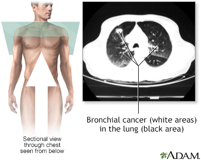
SCLC is the most aggressive form of lung cancer. It usually starts in the breathing tubes (bronchi) in the center of the chest. Although the cancer cells are small, they grow very quickly and create large tumors. These tumors often spread rapidly (metastasize) to other parts of the body, including the brain, liver, and bone.
Symptoms
Symptoms of SCLC include:
- Bloody sputum (phlegm)
Bloody sputum
Coughing up blood is the spitting up of blood or bloody mucus from the lungs and throat (respiratory tract). Hemoptysis is the medical term for cough...
 ImageRead Article Now Book Mark Article
 - Chest pain
Chest pain
Chest pain is discomfort or pain that you feel anywhere along the front of your body between your neck and upper abdomen.
 ImageRead Article Now Book Mark Article
 - Cough
- Loss of appetite
- Shortness of breath
Shortness of breath
Breathing difficulty may involve:Difficult breathing Uncomfortable breathingFeeling like you are not getting enough air
ImageRead Article Now Book Mark Article - Weight loss
Weight loss
Unexplained weight loss is a decrease in body weight, when you did not try to lose the weight on your own. Many people gain and lose weight. Uninten...
Read Article Now Book Mark Article - Wheezing
Wheezing
Wheezing is a high-pitched whistling sound during breathing. It occurs when air moves through narrowed breathing tubes in the lungs.
ImageRead Article Now Book Mark Article
Other symptoms that may occur with this disease, especially in the late stages, include:
- Facial swelling
- Fever
- Hoarseness or changing voice
- Swallowing difficulty
Swallowing difficulty
Difficulty with swallowing is the feeling that food or liquid is stuck in the throat or at any point before the food enters the stomach. This proble...
 ImageRead Article Now Book Mark Article
 - Weakness

Exams and Tests
Your health care provider will perform a physical exam and ask about your medical history. You will be asked whether you smoke, and if so, how much and for how long.
When listening to your chest with a stethoscope, your provider may hear fluid around the lungs or areas where the lung has partially collapsed. Each of these findings may suggest cancer.
SCLC has usually spread to other parts of your body by the time it is diagnosed.
Tests that may be performed include:
- Bone scan
Bone scan
A bone scan is an imaging test used to diagnose bone diseases and find out how severe they are.
 ImageRead Article Now Book Mark Article
 - Chest x-ray
Chest x-ray
A chest x-ray is an x-ray of the chest, lungs, heart, large arteries, ribs, and diaphragm.
 ImageRead Article Now Book Mark Article
 - Complete blood count (CBC)
CBC
A complete blood count (CBC) test measures the following:The number of white blood cells (WBC count)The number of red blood cells (RBC count)The numb...
 ImageRead Article Now Book Mark Article
 - CT scan
CT scan
A chest CT (computed tomography) scan is an imaging method that uses x-rays to create cross-sectional pictures of the chest and upper abdomen....
 ImageRead Article Now Book Mark Article
 - Liver function tests
- MRI scan
MRI scan
A chest MRI (magnetic resonance imaging) scan is an imaging test that uses powerful magnetic fields and radio waves to create pictures of the chest (...
 ImageRead Article Now Book Mark Article
 - Positron emission tomography (PET) scan
- Sputum cytology test (to look for cancer cells), seldom done today
- Thoracentesis (removal of fluid from the chest cavity around the lungs)
In most cases, a piece of tissue is removed from your lungs or other areas to be examined under a microscope. This is called a biopsy. There are several ways to do a biopsy:
- Bronchoscopy combined with biopsy
Bronchoscopy
Bronchoscopy is a test to view the airways and diagnose lung disease. It may also be used during the treatment of some lung conditions.
 ImageRead Article Now Book Mark Article
 - CT scan-directed needle biopsy
CT scan-directed needle biopsy
A lung needle biopsy is a method to remove a piece of lung tissue for examination. If it is done through the wall of your chest, it is called a tran...
 ImageRead Article Now Book Mark Article
 - Endoscopic esophageal or bronchial ultrasound with biopsy
- Mediastinoscopy with biopsy
- Open lung biopsy
- Pleural biopsy
- Video-assisted thoracoscopy
Video-assisted thoracoscopy
Lung surgery is surgery done to repair or remove lung tissue. There are many common lung surgeries, including:Biopsy of an unknown growth in or arou...
 ImageRead Article Now Book Mark Article
Usually, if a biopsy shows cancer, more imaging tests are done to find out the stage of the cancer. Stage means how far it has spread. SCLC is classified as either:
- Limited -- Cancer is only in the chest and can be treated with radiation therapy.
- Extensive -- Cancer has spread outside the area that can be covered by radiation therapy.
Treatment
Because SCLC spreads quickly throughout the body, treatment will include cancer-killing medicines (chemotherapy), which are usually given through a vein (by IV).
Treatment with chemotherapy and immunotherapy, and possibly radiation, may be done for people with SCLC that has spread throughout the body (extensive). In this case, the treatment only helps relieve symptoms and prolongs life, but does not cure the disease.
Radiation therapy can be used with chemotherapy if the disease is confined to one area within the chest (limited).
Radiation therapy uses powerful x-rays or other forms of radiation to kill cancer cells.
Radiation may be used to:
- Treat the cancer, along with chemotherapy, if surgery is not possible.
- Help relieve symptoms caused by the cancer, such as breathing problems and swelling.
- Help relieve cancer pain when the cancer has spread to the bones.
Often, SCLC may have already spread to the brain. This can occur even when there are no symptoms or other signs of cancer in the brain. As a result, some people with smaller cancers, or who had a good response in their first round of chemotherapy, may receive radiation therapy to the brain. This therapy, called prophylactic cranial irradiation, is done to prevent growth of the cancer in the brain.
Surgery helps very few people with SCLC because the disease has often spread by the time it is diagnosed. Surgery may be done when there is only one tumor that has not spread. If surgery is done, chemotherapy or radiation therapy is still needed.
Support Groups
You can ease the stress of illness by joining a cancer support group. Sharing with others who have common experiences and problems can help you not feel alone.
Cancer support group
The following organizations are good resources for information on cancer:American Cancer Society. Support and online communities. www. cancer. org/...
Read Article Now Book Mark ArticleOutlook (Prognosis)
How well you do depends on how much the lung cancer has spread. SCLC is very deadly. Not many people with this type of cancer are still alive 5 years after diagnosis.
Treatment can often prolong life to more than 12 months, even when the cancer has spread.
In rare cases, if SCLC is diagnosed early, treatment may result in a long-term cure.
When to Contact a Medical Professional
Contact your provider if you have symptoms of lung cancer, particularly if you smoke.
Prevention
If you smoke, now is the time to quit. If you are having trouble quitting, talk with your provider. There are many methods to help you quit, from support groups to prescription medicines. Also try to avoid secondhand smoke.
Trouble quitting
There are many ways to quit smoking. There are also resources to help you. Family members, friends, and co-workers may be supportive. But to be su...

If you smoke or used to smoke, talk with your provider about getting screened for lung cancer. To get screened, you need to have a CT scan of the chest.
Reviewed By
Warren Brenner, MD, Oncologist, Lynn Cancer Institute, Boca Raton, FL. Review provided by VeriMed Healthcare Network. Also reviewed by David C. Dugdale, MD, Medical Director, Brenda Conaway, Editorial Director, and the A.D.A.M. Editorial team.
Araujo LH, Horn L, Merritt RE, Shilo K, Xu-Welliver M, Carbone DP. Cancer of the lung: non-small cell lung cancer and small cell lung cancer. In: Niederhuber JE, Armitage JO, Kastan MB, Doroshow JH, Tepper JE, eds. Abeloff's Clinical Oncology. 6th ed. Philadelphia, PA: Elsevier; 2020:chap 69.
National Comprehensive Cancer Network website. NCCN clinical practice guidelines in oncology: Small cell lung cancer. Version 4. 2025. www.nccn.org/professionals/physician_gls/pdf/sclc.pdf. Updated January 13, 2025. Accessed July 18, 2025.
National Cancer Institute website. Small cell lung cancer treatment (PDQ) - health professional version. www.cancer.gov/types/lung/hp/small-cell-lung-treatment-pdq. Updated May 14, 2025. Accessed July 18, 2025.
Rivera MP, Mody GN, Weiner AA. Lung cancer: treatment. In: Broaddus VC, Ernst JD, Talmadge EK, et al, eds. Murray and Nadel's Textbook of Respiratory Medicine. 7th ed. Philadelphia, PA: Elsevier; 2022:chap 77.