Uvea
Vascular tunic
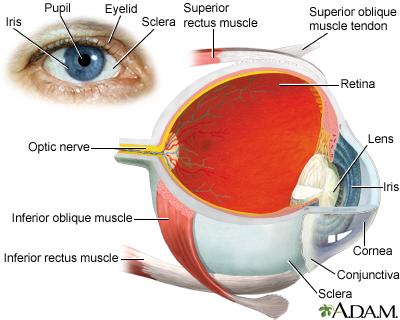
The uvea is the middle layer of the eye. It lies beneath the white part of the eye (the sclera). It is made of the iris, ciliary body, and choroid. These structures control many eye functions, including adjusting to different levels of light or distances of objects. Inflammation of one or more of these structures is called uveitis.
Images
Related Information
ScleraIris
Ciliary body
Choroid
References
Evans M. Anatomy of the uvea. In: Yanoff M, Duker JS, eds. Ophthalmology. 6th ed. Philadelphia, PA: Elsevier; 2023:chap 7.1.
Taber's Cyclopedic Medical Dictionary. 24th ed. F.A. Davis Company; 2021. www.tabers.com/tabersonline/view/Tabers-Dictionary/770909/0/uvea. Accessed July 8, 2025.
BACK TO TOPReview Date: 11/1/2023
Reviewed By: Linda J. Vorvick, MD, Clinical Professor, Department of Family Medicine, UW Medicine, School of Medicine, University of Washington, Seattle, WA. Also reviewed by David C. Dugdale, MD, Medical Director, Brenda Conaway, Editorial Director, and the A.D.A.M. Editorial team. Editorial update 07/08/2025.

Health Content Provider
06/01/2028
|
A.D.A.M., Inc. is certified by URAC, for Health Content Provider (www.urac.org). URAC's certification program is an independent audit to verify that A.D.A.M. follows rigorous standards of quality and accountability. A.D.A.M. is among the first to achieve this important distinction for online health information and services. Learn more about A.D.A.M.'s editorial policy, editorial process and privacy policy. |
The information provided herein should not be used during any medical emergency or for the diagnosis or treatment of any medical condition. A licensed medical professional should be consulted for diagnosis and treatment of any and all medical conditions. Links to other sites are provided for information only -- they do not constitute endorsements of those other sites. © 1997- 2026 A.D.A.M., a business unit of Ebix, Inc. Any duplication or distribution of the information contained herein is strictly prohibited.
 All rights reserved.