Electroretinography

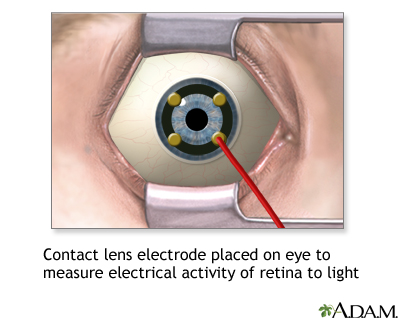
Contact lens electrode on eye
Schedule an Appointment
Definition
Electroretinography is a test to measure the electrical response of the eye's light-sensitive cells, called rods and cones. These cells are part of the retina (the back part of the eye).
How the Test is Performed
While you are in a sitting position, the health care provider places numbing drops into your eyes, so you will not have any discomfort during the test. Your eyes are held open with a small device called a speculum. An electrical sensor (electrode) is placed on each eye.
The electrode measures the electrical activity of the retina in response to light. A light flashes, and the electrical response travels from the electrode to a TV-like screen, where it can be viewed and recorded. The normal response pattern has waves called A and B.
The provider will take the readings in normal room light and then again in the dark, after allowing 20 minutes for your eyes to adjust.
How to Prepare for the Test
No special preparation is necessary for this test.
How the Test will Feel
The probes that rest on your eye may feel a little scratchy. The test takes about 1 hour to perform.
Why the Test is Performed
This test is done to detect disorders of the retina. It is also useful for determining if retinal surgery is recommended.
Normal Results
Normal test results will show a normal A and B pattern in response to each flash.
What Abnormal Results Mean
The following conditions may cause abnormal results:
- Arteriosclerosis with damage to the retina
- Congenital night blindness
- Congenital retinoschisis (splitting of the retinal layers)
- Giant cell arteritis
- Medicines (chloroquine, hydroxychloroquine)
- Mucopolysaccharidosis
- Retinal detachment
- Rod-cone dystrophy (retinitis pigmentosa)
- Trauma
- Vitamin A deficiency
Risks
The cornea may get a temporary scratch on the surface from the electrode. Otherwise, there are no risks with this procedure.
Considerations
You should not rub your eyes for an hour after the test, as this could injure the cornea. Your provider will talk to you about the results of the test and what they mean for you.
References
Baloh RW, Jen JC. Neuro-ophthalmology. In: Goldman L, Cooney KA, eds. Goldman-Cecil Medicine. 27th ed. Philadelphia, PA: Elsevier; 2024:chap 392.
Reichel E, Klein K, Richard A. Retinal electrophysiology. In: Yanoff M, Duker JS, eds. Ophthalmology. 6th ed. Philadelphia, PA: Elsevier; 2023:chap 6.7.
Tsang SH, Holder GE. Clinical electrophysiology. In: Sadda SVR, Sarraf D, Freund B, et al, eds. Ryan's Retina. 7th ed. Philadelphia, PA: Elsevier; 2023:chap 9.

Review Date: 8/5/2024
Reviewed By: Franklin W. Lusby, MD, Ophthalmologist, Lusby Vision Institute, La Jolla, CA. Also reviewed by David C. Dugdale, MD, Medical Director, Brenda Conaway, Editorial Director, and the A.D.A.M. Editorial team.
The information provided herein should not be used during any medical emergency or for the diagnosis or treatment of any medical condition. A licensed medical professional should be consulted for diagnosis and treatment of any and all medical conditions. Links to other sites are provided for information only -- they do not constitute endorsements of those other sites. No warranty of any kind, either expressed or implied, is made as to the accuracy, reliability, timeliness, or correctness of any translations made by a third-party service of the information provided herein into any other language.
© 1997-
A.D.A.M., a business unit of Ebix, Inc. Any duplication or distribution of the information contained herein is strictly prohibited.
All content on this site including text, images, graphics, audio, video, data, metadata, and compilations is protected by copyright and other intellectual property laws. You may view the content for personal, noncommercial use. Any other use requires prior written consent from Ebix. You may not copy, reproduce, distribute, transmit, display, publish, reverse-engineer, adapt, modify, store beyond ordinary browser caching, index, mine, scrape, or create derivative works from this content. You may not use automated tools to access or extract content, including to create embeddings, vectors, datasets, or indexes for retrieval systems. Use of any content for training, fine-tuning, calibrating, testing, evaluating, or improving AI systems of any kind is prohibited without express written consent. This includes large language models, machine learning models, neural networks, generative systems, retrieval-augmented systems, and any software that ingests content to produce outputs. Any unauthorized use of the content including AI-related use is a violation of our rights and may result in legal action, damages, and statutory penalties to the fullest extent permitted by law. Ebix reserves the right to enforce its rights through legal, technological, and contractual measures.