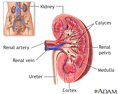


Renal scan
A renal scan is a nuclear medicine exam in which a small amount of radioactive material (radioisotope) is used to measure the function of the kidneys.
How the Test is Performed
The specific type of scan may vary. This article provides a general overview.
A renal scan is similar to a renal perfusion scintiscan. It may be done along with that test.
You will be asked to lie on the scanner table. The health care provider will place a tight band or blood pressure cuff on your upper arm. This creates pressure and helps your arm veins become bigger. A small amount of radioisotope is injected into a vein. The specific radioisotope used may vary, depending on what is being studied.
The cuff or band on the upper arm is removed, and the radioactive material moves through your blood. The kidneys are scanned a short time later. Several images are taken, each lasting 1 or 2 seconds. The scan takes about 30 minutes to 1 hour in total.
A computer reviews the images and provides detailed information about how your kidney works. For example, it can tell your provider how much blood the kidney filters over time. A diuretic medicine (water pill) may also be injected during the test. This helps speed up the passage of radioisotope through your kidneys.
You should be able to go home after the scan. You may be asked to drink plenty of fluids and urinate often to help remove the radioactive material from the body.
How to Prepare for the Test
Tell your provider if you take any nonsteroidal anti-inflammatory drugs (NSAIDs) or blood pressure medicines. These medicines might affect the test.
You may be asked to drink additional fluids before the scan.
How the Test will Feel
Some people feel discomfort when the needle is placed into the vein. However, you will not feel the radioactive material. The scanning table may be hard and cold. You will need to lie still during the scan. You may feel an increased urge to urinate near the end of the test.
Why the Test is Performed
A renal scan tells your provider how your kidneys work. It also shows their size, position, and shape. It may be done if:
- You cannot have other x-rays using contrast (dye) material because you are sensitive or allergic to them, or you have reduced kidney function
- You have had a kidney transplant and your provider wants to check how well the kidney is working and look for signs of rejection
- You have high blood pressure and your provider wants to see how well your kidneys are working
- Your provider needs to confirm if a kidney that looks swollen or blocked on another imaging test is losing function
What Abnormal Results Mean
Abnormal results are a sign of reduced kidney function. This may be due to:
- Acute or chronic kidney failure
- Chronic kidney infection (pyelonephritis)
- Complications of a kidney transplant
Kidney transplant
A kidney transplant is surgery to place a healthy kidney into a person with kidney failure.
 ImageRead Article Now Book Mark Article
 - Glomerulonephritis
- Hydronephrosis
- Injury of the kidney and ureter
- Narrowing or blockage of the arteries that carry blood to the kidney
- Obstructive uropathy
Risks
There is a slight amount of radiation from the radioisotope. Most of this radiation exposure occurs to the kidneys and bladder. Almost all radiation is gone from the body in 24 hours. Caution is advised if you are pregnant or breastfeeding.
Very rarely, a person will have an allergic reaction to the radioisotope, which may include severe anaphylaxis.
Reviewed By
Kelly L. Stratton, MD, FACS, Associate Professor, Department of Urology, University of Oklahoma Health Sciences Center, Oklahoma City, OK. Also reviewed by David C. Dugdale, MD, Medical Director, Brenda Conaway, Editorial Director, and the A.D.A.M. Editorial team.
Duddalwar VA, Jadvar H, Palmer SL. Diagnostic kidney imaging. In: Yu ASL, Chertow GM, Luyckx VA, Marsden PA, Skorecki K, Taal MW, eds. Brenner and Rector's The Kidney. 11th ed. Philadelphia, PA: Elsevier; 2020:chap 25.
Shukla AR, Srinivasan AK. Posterior urethral valves. In: Partin AW, Dmochowski RR, Kavoussi LR, Peters CA, eds. Campbell-Walsh-Wein Urology. 12th ed. Philadelphia, PA: Elsevier; 2021:chap 33.
Wymer DTG, Wymer DC. Radiologic and nuclear imaging in nephrology. In: Johnson RJ, Floege J, Tonelli M, eds. Comprehensive Clinical Nephrology. 7th ed. Philadelphia, PA: Elsevier; 2024:chap 6.