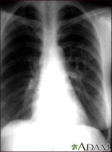


Chest x-ray
A chest x-ray is an x-ray of the chest, lungs, heart, large arteries, ribs, and diaphragm.
x-ray
X-rays are a type of electromagnetic radiation, just like visible light. An x-ray machine sends individual x-ray waves through the body. The images...

How the Test is Performed
You stand in front of the x-ray machine. You will be told to take a breath in and hold it when the x-ray is taken.
Two images are usually taken. You will first need to stand facing the machine, and then sideways.
How to Prepare for the Test
Tell your health care provider if you are pregnant. Chest x-rays are generally avoided during pregnancy, and special precautions are taken if they are needed.
How the Test will Feel
There is no discomfort. The imaging plate may feel cold.
Why the Test is Performed
Your provider may order a chest x-ray if you have any of the following symptoms:
- A persistent cough
- Chest pain from a chest injury (with a possible rib fracture or lung complication) or from heart problems
Chest pain
Chest pain is discomfort or pain that you feel anywhere along the front of your body between your neck and upper abdomen.
 ImageRead Article Now Book Mark Article
 - Coughing up blood
Coughing up blood
Coughing up blood is the spitting up of blood or bloody mucus from the lungs and throat (respiratory tract). Hemoptysis is the medical term for cough...
 ImageRead Article Now Book Mark Article
 - Difficulty breathing
Difficulty breathing
Breathing difficulty may involve:Difficult breathing Uncomfortable breathingFeeling like you are not getting enough air
 ImageRead Article Now Book Mark Article
 - Fever
It may also be done if you have signs of tuberculosis, lung cancer, or other chest or lung diseases.
Lung cancer
Lung cancer is cancer that starts in the lungs. The lungs are located in the chest. When you breathe, air goes through your nose, down your windpipe...
Lung diseases
Lung disease is any problem in the lungs that prevents the lungs from working properly. There are three main types of lung disease:Airway diseases -...

A serial chest x-ray is one that is repeated. It may be done to monitor changes found on a past chest x-ray.
What Abnormal Results Mean
Abnormal results may be due to many things, including:
In the lungs:
- Collapsed lung
- Collection of fluid around the lung
- Lung tumor (noncancerous or cancerous)
- Malformation of the blood vessels
- Pneumonia
- Scarring of lung tissue
- Tuberculosis
- Atelectasis
In the heart:
- Problems with the size, position or shape of the heart
- Problems with the position, size and shape of the large arteries
- Evidence of heart failure
In the bones:
- Fractures or other problems of the ribs and spine
- Osteoporosis
Osteoporosis
Osteoporosis is a disease in which bones become fragile and more likely to break (fracture).
 ImageRead Article Now Book Mark Article
In the mediastinum (middle part of the chest):
- Enlargement, which might be related to infection or tumor
Risks
There is low radiation exposure. X-rays are monitored and regulated to provide the minimum amount of radiation exposure needed to produce the image. Most experts feel that the benefits outweigh the risks. Pregnant women and children are more sensitive to the risks of x-rays.
Reviewed By
Allen J. Blaivas, DO, Division of Pulmonary, Critical Care, and Sleep Medicine, VA New Jersey Health Care System, Clinical Assistant Professor, Rutgers New Jersey Medical School, East Orange, NJ. Review provided by VeriMed Healthcare Network. Also reviewed by David C. Dugdale, MD, Medical Director, Brenda Conaway, Editorial Director, and the A.D.A.M. Editorial team.
Felker GM, Teerlink JR. Diagnosis and management of acute heart failure. In: Libby P, Bonow RO, Mann DL, Tomaselli GF, Bhatt DL, Solomon SD, eds. Braunwald's Heart Disease: A Textbook of Cardiovascular Medicine. 12th ed. Philadelphia, PA: Elsevier; 2022:chap 49.
Jokerst CE, Gotway MB. Thoracic radiology: noninvasive diagnostic imaging. In: Broaddus VC, Ernst JD, King TE, et al, eds. Murray and Nadel's Textbook of Respiratory Medicine. 7th ed. Philadelphia, PA: Elsevier; 2022:chap 20.
Nair A, Barnett JL, Semple TR. Current status of thoracic imaging. In: Adam A, Dixon AK, Gillard JH, Schaefer-Prokop CM, eds. Grainger & Allison's Diagnostic Radiology. 7th ed. Philadelphia, PA: Elsevier; 2021:chap 1.
Disclaimer
© 1997- A.D.A.M., a business unit of Ebix, Inc. Any duplication or distribution of the information contained herein is strictly prohibited.