Surgeries, Tests And Treatments
More <
bookmarks-menu

Thoracic spine x-ray

Show Alternative Names
Vertebral radiography
X-ray - spine
Thoracic x-ray
Spine x-ray
Thoracic spine films
Back films

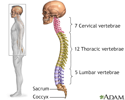
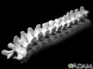
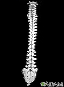
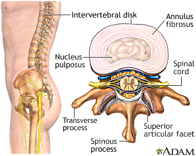
A thoracic spine x-ray is an x-ray of the 12 chest (thoracic) bones (vertebrae) of the spine. The vertebrae are separated by flat pads of cartilage called disks that provide a cushion between the bones.

How the Test is Performed

The test is done in a hospital radiology department or in your health care provider's office. You will lie on the x-ray table in different positions. If the x-ray is checking for an injury, care will be taken to prevent further injury.

The x-ray machine will be moved over the thoracic area of the spine. You will hold your breath as the picture is taken so that the picture will not be blurry. Usually, 2 or 3 x-ray views are needed.

How to Prepare for the Test

Tell your provider if you are pregnant. Also tell your provider if you have had surgery in your chest, abdomen, or pelvis.

Remove all jewelry and accessories.

How the Test will Feel

The test causes no discomfort. The table may be cold.

Why the Test is Performed

The x-ray helps evaluate:

What Abnormal Results Mean

The test can detect:

Risks

There is low radiation exposure. X-rays are monitored and regulated to provide the minimum amount of radiation exposure needed to produce the image. Most experts feel that the risk is low compared with the benefits.

Pregnant women and children are more sensitive to the risks of x-rays.

Considerations

The x-ray will not detect problems in the muscles, nerves, and other soft tissues, because these problems cannot be seen well on an x-ray.

Review Date: 9/2/2025

Reviewed By

C. Benjamin Ma, MD, Professor, Chief, Sports Medicine and Shoulder Service, UCSF Department of Orthopaedic Surgery, San Francisco, CA. Also reviewed by David C. Dugdale, MD, Medical Director, Brenda Conaway, Editorial Director, and the A.D.A.M. Editorial team.

References

Preston-Suni K, Kaji AH. Spinal trauma. In: Walls RM, ed. Rosen's Emergency Medicine: Concepts and Clinical Practice. 10th ed. Philadelphia, PA: Elsevier; 2023:chap 35.

Van Thielen T, van den Hauwe L, Van Goethem JW, Parizel PM. Current status of imaging of the spine and anatomical features. In: Adam A, Dixon AK, Gillard JH, Schaefer-Prokop CM, eds. Grainger & Allison's Diagnostic Radiology: A Textbook of Medical Imaging. 7th ed. Philadelphia, PA: Elsevier; 2021:chap 47.

Disclaimer

The information provided herein should not be used during any medical emergency or for the diagnosis or treatment of any medical condition. A licensed medical professional should be consulted for diagnosis and treatment of any and all medical conditions. Links to other sites are provided for information only -- they do not constitute endorsements of those other sites. No warranty of any kind, either expressed or implied, is made as to the accuracy, reliability, timeliness, or correctness of any translations made by a third-party service of the information provided herein into any other language.

© 1997- A.D.A.M., a business unit of Ebix, Inc. Any duplication or distribution of the information contained herein is strictly prohibited.

All content on this site including text, images, graphics, audio, video, data, metadata, and compilations is protected by copyright and other intellectual property laws. You may view the content for personal, noncommercial use. Any other use requires prior written consent from Ebix. You may not copy, reproduce, distribute, transmit, display, publish, reverse-engineer, adapt, modify, store beyond ordinary browser caching, index, mine, scrape, or create derivative works from this content. You may not use automated tools to access or extract content, including to create embeddings, vectors, datasets, or indexes for retrieval systems. Use of any content for training, fine-tuning, calibrating, testing, evaluating, or improving AI systems of any kind is prohibited without express written consent. This includes large language models, machine learning models, neural networks, generative systems, retrieval-augmented systems, and any software that ingests content to produce outputs. Any unauthorized use of the content including AI-related use is a violation of our rights and may result in legal action, damages, and statutory penalties to the fullest extent permitted by law. Ebix reserves the right to enforce its rights through legal, technological, and contractual measures.

All
Videos
Images
Skeletal spine - Illustration Thumbnail

Skeletal spine

The spine is divided into several sections. The cervical vertebrae make up the neck. The thoracic vertebrae comprise the chest section and have ribs attached. The lumbar vertebrae are the remaining vertebrae below the last thoracic bone and the top of the sacrum. The sacral vertebrae are caged within the bones of the pelvis, and the coccyx represents the terminal vertebrae or vestigial tail.

Illustration

Vertebra, thoracic (mid back) - Illustration Thumbnail

Vertebra, thoracic (mid back)

These are twelve vertebra of the mid back. The last vertebra (on the left side of the picture) attaches to the lumbar (lower) spine, and the top vertebra (on the right) attaches to the cervical (neck) section of the back. The vertebra are broader and stronger than the cervical bones. This allows them to absorb the added pressure applied to the mid back, but they remain a common sight of injury. The vertebra are numbered from one to twelve and labeled T1, T2, T3, et cetera, from the upper most bones to the lowest.

Illustration

Vertebral column - Illustration Thumbnail

Vertebral column

This is the spine and the sacrum with the cervical (neck), thoracic (mid-back), and lumbar (lower back) vertebra. Notice how the appearance of the vertebra change as you look down the spine. The change in shape and size reflect the different functions of the neck, mid-back, and lower back.

Illustration

Intervertebral disk - Illustration Thumbnail

Intervertebral disk

The vertebral column is made up of 26 bones that provide axial support to the trunk. The vertebral column provides protection to the spinal cord that runs through its central cavity. Between each vertebra is an intervertebral disk. The disks are filled with a gelatinous substance, called the nucleus pulposus, which provides cushioning to the spinal column. The annulus fibrosus is a fibrocartilaginous ring that surrounds the nucleus pulposus, which keeps the nucleus pulposus intact when forces are applied to the spinal column. The intervertebral disks allow the vertebral column to be flexible and act as shock absorbers during everyday activities such as walking, running and jumping.

Illustration

Anterior skeletal anatomy - Illustration Thumbnail

Anterior skeletal anatomy

The skeleton is made up of 206 bones in the adult and contributes to the form and shape of the body. The skeleton has several important functions for the body. The bones of the skeleton provide support for the soft tissues. For example, the rib cage supports the thoracic wall. Most muscles of the body are attached to bones which act as levers to allow movement of body parts. The bones of the skeleton also serve as a reservoir for minerals, such as calcium and phosphate. Finally, most of the blood cell formation takes places within the marrow of certain bones.

Illustration


The information provided herein should not be used during any medical emergency or for the diagnosis or treatment of any medical condition. A licensed medical professional should be consulted for diagnosis and treatment of any and all medical conditions. Links to other sites are provided for information only -- they do not constitute endorsements of those other sites. No warranty of any kind, either expressed or implied, is made as to the accuracy, reliability, timeliness, or correctness of any translations made by a third-party service of the information provided herein into any other language.

© 1997- A.D.A.M., a business unit of Ebix, Inc. Any duplication or distribution of the information contained herein is strictly prohibited.

All content on this site including text, images, graphics, audio, video, data, metadata, and compilations is protected by copyright and other intellectual property laws. You may view the content for personal, noncommercial use. Any other use requires prior written consent from Ebix. You may not copy, reproduce, distribute, transmit, display, publish, reverse-engineer, adapt, modify, store beyond ordinary browser caching, index, mine, scrape, or create derivative works from this content. You may not use automated tools to access or extract content, including to create embeddings, vectors, datasets, or indexes for retrieval systems. Use of any content for training, fine-tuning, calibrating, testing, evaluating, or improving AI systems of any kind is prohibited without express written consent. This includes large language models, machine learning models, neural networks, generative systems, retrieval-augmented systems, and any software that ingests content to produce outputs. Any unauthorized use of the content including AI-related use is a violation of our rights and may result in legal action, damages, and statutory penalties to the fullest extent permitted by law. Ebix reserves the right to enforce its rights through legal, technological, and contractual measures.
© 1997- adam.com All rights reserved.