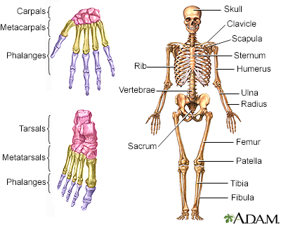


Bone x-ray
A bone x-ray is an imaging test to look at the bones.
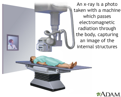
x-ray
X-rays are a type of electromagnetic radiation, just like visible light. An x-ray machine sends individual x-ray waves through the body. The images...

How the Test is Performed
The test is done in a hospital radiology department or in your health care provider's office by an x-ray technician. For the test, you will position the bone to be x-rayed on the table or in other positions, depending on the body part. Pictures are then taken, and the bone is repositioned for different views.
How to Prepare for the Test
Tell your provider if you are pregnant. You must remove all jewelry for the x-ray.
How the Test will Feel
The x-rays are painless. Changing position for getting different views of the bone may be uncomfortable.
Why the Test is Performed
A bone x-ray is used to look for injuries or conditions affecting the bone.
What Abnormal Results Mean
Abnormal findings include:
- Fractures or broken bone
Fractures or broken bone
If more pressure is put on a bone than it can stand, it will split or break. A break of any size is called a fracture. If the broken bone punctures...
ImageRead Article Now Book Mark Article - Bone tumors
- Degenerative bone conditions
- Osteomyelitis (bone infection)
- Arthritis
Additional conditions under which the test may be performed:
- Multiple myeloma
- Osgood-Schlatter disease
- Osteogenesis imperfecta
- Osteomalacia
- Paget's disease
- Primary hyperparathyroidism
- Rickets
Risks
There is low radiation exposure. X-ray machines are set to provide the smallest amount of radiation exposure needed to produce the image. Most experts feel that the risk is low compared with the benefits.
Children and the fetuses of pregnant women are more sensitive to the risks of the x-ray. A protective shield may be worn over areas not being scanned.
Reviewed By
Jason Levy, MD, FSIR, Northside Radiology Associates, Atlanta, GA. Also reviewed by David C. Dugdale, MD, Medical Director, Brenda Conaway, Editorial Director, and the A.D.A.M. Editorial team.
Contreras F, Perez J, Jose J. Imaging overview. In: Miller MD, Thompson SR. eds. DeLee, Drez, & Miller's Orthopaedic Sports Medicine. 5th ed. Philadelphia, PA: Elsevier; 2020:chap 7.
Kapoor G, Toms AP. Current status of imaging of the musculoskeletal system. In: Adam A, Dixon AK, Gillard JH, Schaefer-Prokop CM, eds. Grainger & Allison's Diagnostic Radiology: A Textbook of Medical Imaging. 7th ed. Philadelphia, PA: Elsevier; 2021:chap 38.
Disclaimer
© 1997- A.D.A.M., a business unit of Ebix, Inc. Any duplication or distribution of the information contained herein is strictly prohibited.