Multimedia Gallery


CSF oligoclonal banding - series
CSF oligoclonal banding - series
The cerebrospinal fluid (CSF) serves to supply nutrients to the central nervous system (CNS) and collect waste products, as well as provide lubrication.
CSF oligoclonal banding - series
CSF oligoclonal banding - series
The cerebrospinal fluid (CSF) serves to supply nutrients to the central nervous system (CNS) and collect waste products, as well as provide lubrication.
CSF oligoclonal banding - series
Indications
Why the test is performed.
This test helps confirm the diagnosis of multiple sclerosis (MS).
The laboratory procedure is called CSF electrophoresis, which is a method used to study the levels of protein in the cerebrospinal fluid (CSF).
In electrophoresis, a CSF sample is applied to a gel (solid medium allowing the movement of proteins), and a voltage is applied. The proteins migrate along the gel based on their charge (roughly on their size). The gel is stained, and significant amounts of similar proteins will cause a visible "band" to be present. The term oligoclonal bands refers the presence in CSF of two or more protein bands of a specific immunoglobin (IgG) that have greater intensity than in the concurrent serum sample. This pattern of banding is seen in patients with MS, and other conditions.
CSF oligoclonal banding - series
Indications
Why the test is performed.This test helps confirm the diagnosis of multiple sclerosis (MS).The laboratory procedure is called CSF electrophoresis, wh...
CSF oligoclonal banding - series
Procedure, part 1
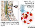
A sample of the CSF will be taken from the lumbar area of the spine. This is called a lumbar puncture. How the test will feel: The position used during lumbar puncture may be uncomfortable, but you must remain in the curled position to avoid moving the needle and possibly injuring the spinal cord. There also may be some discomfort with the needle prick and the insertion of the lumbar puncture needle. When the fluid is withdrawn, there may be a feeling of pressure.
Risks of lumbar puncture include:
- Allergic reaction to the anesthetic.
- Discomfort during the test.
- Headache after the test.
- Bleeding into the spinal canal.
- Brain herniation (if performed on a patient with increased intracranial pressure), which can result in brain damage and/or death.
- Damage to the spinal cord (particularly ithe patient moves during the test).
CSF oligoclonal banding - series
Procedure, part 1
A sample of the CSF will be taken from the lumbar area of the spine. This is called a lumbar puncture. How the test will feel: The position used duri...
CSF oligoclonal banding - series
Procedure, part 2
A blood sample will also be drawn.
CSF oligoclonal banding - series
Procedure, part 2
A blood sample will also be drawn.
CSF oligoclonal banding - series
Procedure, part 3
Both the samples are added to a gel, which is then filtered through a cartridge. The gel separates the proteins in each sample and the lab looks for oligoclonal banding.
If results are normal one or less bandings will be found in the CSF.
If results are abnormal there are 2 or more bandings found in the CSF and not in the blood serum. This may indicate multiple sclerosis (MS). CSF oligoclonal bands are found in 83% to 94% of patients with definite MS. Other causes of oligoclonal banding in CSF include encephalitis, meningitis, Guillain-Barre syndrome, polyneuritis, headache, and other conditions.
CSF oligoclonal banding - series
Procedure, part 3
Both the samples are added to a gel, which is then filtered through a cartridge. The gel separates the proteins in each sample and the lab looks for ...
Review Date: 4/16/2025
Reviewed By: Joseph V. Campellone, MD, Department of Neurology, Cooper Medical School at Rowan University, Camden, NJ. Review provided by VeriMed Healthcare Network. Also reviewed by David C. Dugdale, MD, Medical Director, Brenda Conaway, Editorial Director, and the A.D.A.M. Editorial team.
© 1997- A.D.A.M., a business unit of Ebix, Inc. Any duplication or distribution of the information contained herein is strictly prohibited.
The cerebrospinal fluid (CSF) serves to supply nutrients to the central nervous system (CNS) and collect waste products, as well as provide lubrication.
Why the test is performed.
This test helps confirm the diagnosis of multiple sclerosis (MS).
The laboratory procedure is called CSF electrophoresis, which is a method used to study the levels of protein in the cerebrospinal fluid (CSF).
In electrophoresis, a CSF sample is applied to a gel (solid medium allowing the movement of proteins), and a voltage is applied. The proteins migrate along the gel based on their charge (roughly on their size). The gel is stained, and significant amounts of similar proteins will cause a visible "band" to be present. The term oligoclonal bands refers the presence in CSF of two or more protein bands of a specific immunoglobin (IgG) that have greater intensity than in the concurrent serum sample. This pattern of banding is seen in patients with MS, and other conditions.
A sample of the CSF will be taken from the lumbar area of the spine. This is called a lumbar puncture. How the test will feel: The position used during lumbar puncture may be uncomfortable, but you must remain in the curled position to avoid moving the needle and possibly injuring the spinal cord. There also may be some discomfort with the needle prick and the insertion of the lumbar puncture needle. When the fluid is withdrawn, there may be a feeling of pressure.
Risks of lumbar puncture include:
- Allergic reaction to the anesthetic.
- Discomfort during the test.
- Headache after the test.
- Bleeding into the spinal canal.
- Brain herniation (if performed on a patient with increased intracranial pressure), which can result in brain damage and/or death.
- Damage to the spinal cord (particularly ithe patient moves during the test).
A blood sample will also be drawn.
Both the samples are added to a gel, which is then filtered through a cartridge. The gel separates the proteins in each sample and the lab looks for oligoclonal banding.
If results are normal one or less bandings will be found in the CSF.
If results are abnormal there are 2 or more bandings found in the CSF and not in the blood serum. This may indicate multiple sclerosis (MS). CSF oligoclonal bands are found in 83% to 94% of patients with definite MS. Other causes of oligoclonal banding in CSF include encephalitis, meningitis, Guillain-Barre syndrome, polyneuritis, headache, and other conditions.


Animations
- Alzheimer disease
- Alzheimer disease
- Asperger syndrome
- Atherosclerosis
- Athetosis resulting from ba...
- Attention deficit hyperacti...
- Autism spectrum disorders
- Bladder function - neurolog...
- Brain components
- Cerebral aneurysm
- Cerebral palsy
- Cholesterol and triglycerid...
- Cluster headache
- Concussion
- Concussion
- Diabetes
- Epilepsy
- Epinephrine and exercise
- Erection problems
- Essential hypertension
- Feeling pain
- Hardening of arteries
- Head injury
- Herniated disk
- Herniated nucleus pulposus ...
- How to use a pill cutter
- Hypertension - overview
- Insomnia
- Migraine
- Multiple sclerosis
- Neck pain
- Nerve conduction
- Parkinson disease
- Sciatica
- Seizures
- Shingles
- Sleep disorders
- Spinal stenosis
- Stroke
- Stroke
- Stroke - secondary to cardi...
- Systemic lupus erythematosus
- Tension headache
- Understanding cholesterol r...
- Urinary incontinence
Illustrations
- 15/15 rule
- Active vs. inactive muscle
- Acupuncture
- Adult dermatome
- Alcoholic neuropathy
- Alpha-glucosidase inhibitors
- Alzheimer disease
- Amebic brain abscess
- Amyloidosis of the fingers
- Arterial plaque build-up
- Arterial tear in internal c...
- Arteries of the brain
- Atherosclerosis
- Atherosclerosis of internal...
- Autonomic Nerves
- Balance receptors
- Before and after hematoma repair
- Bicycle helmet - proper usage
- Biguanides
- Blood pressure
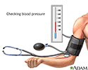
- Blood pressure check
- Blood test
- Blood test
- Bone graft harvest
- Brain
- Brain
- Brain and nervous system
- Brain herniation
- Brain structures
- Brain wave monitor
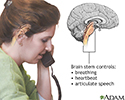
- Brainstem function
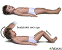
- Brudzinski's sign of meningitis
- Calories and fat per serving
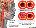
- Carotid dissection
- Carotid duplex
- Carotid stenosis - X-ray of...
- Carotid stenosis - X-ray of...
- Carpal tunnel surgical procedure
- Carpal tunnel syndrome
- Cataract - close-up of the eye
- Cauda equina
- Cause of headaches
- Causes of secondary headache
- Central nervous system
- Central nervous system
- Central nervous system and ...
- Cerebellum - function
- Cerebral aneurysm
- Cerebral aneurysm
- Cerebrospinal fluid leak
- Cervical spondylosis
- Cervical vertebrae
- Cholesterol
- Cholesterol producers
- Circle of Willis
- Common peroneal nerve dysfu...
- Complex carbohydrates
- Compression fracture
- Compression of the median nerve
- Concussion
- Congenital toxoplasmosis
- Copper urine test
- Coronary artery blockage
- Craniotomy for cerebral shunt
- Creutzfeldt-Jakob disease
- Crossed eyes
- CSF cell count
- CSF chemistry
- CSF protein test
- CT scan
- CT scan of the brain
- Damaged axillary nerve
- DASH diet
- Developmental process of at...
- Diabetes and exercise
- Diabetes and nerve damage
- Diabetic blood circulation ...
- Diabetic emergency supplies
- Diabetic retinopathy
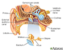
- Ear anatomy
- Effects of age on blood pressure
- Electromyography
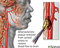
- Endarterectomy
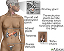
- Endocrine glands
- Enlarged view of atherosclerosis
- Exercise can lower blood pr...
- External and internal eye a...
- Eye
- Eye ultrasound
- Facial drooping
- Femoral nerve damage
- Fibromyalgia
- Fish in diet
- Food and insulin release
- Forward bend test
- Glaucoma
- Glossopharyngeal neuralgia
- Glucose in blood
- Glucose test
- Grand mal seizure
- Gray and white matter of th...
- Haemophilus influenzae organism
- Head injury
- Head trauma
- Headache
- Healthy diet
- Herniated disk repair
- Herniated lumbar disk
- Herniated nucleus pulposus
- Herpes zoster (shingles) - ...
- Herpes zoster (shingles) di...
- Herpes zoster (shingles) on...
- Herpes zoster (shingles) on...
- Herpes zoster (shingles) on...
- Herpes zoster (shingles) on...
- Herpes zoster (shingles) on...
- Herpes zoster (shingles) on...
- Herpes zoster (shingles) on...
- High blood pressure tests
- Hunger center in brain
- Hypothalamus
- Increased intracranial pressure
- Indications of head injury
- Insulin production and diabetes
- Insulin pump
- Insulin pump
- Intervertebral disk
- Intracerebellar hemorrhage ...
- Intracerebral hemorrhage
- Intracranial pressure monitoring
- Irregular sleep
- Kernig's sign of meningitis
- Kyphosis
- Late-stage syphilis
- Left cerebral hemisphere - ...
- Leukoencephalopathy
- Lifestyle changes
- Limbic system
- Lobes of the brain
- Location of whiplash pain
- Low blood sugar symptoms
- Lower leg muscles
- Lumbar puncture (spinal tap)
- Lumbar vertebrae
- Lumbar vertebrae
- Lyme disease
- Lyme disease - Borrelia bur...
- Lyme disease - erythema migrans
- Lyme disease organism - Bor...
- Meninges of the brain
- Meninges of the spine
- Migraine cause
- Migraine headache
- Monitoring blood pressure
- Motor nerves
- MRI of the brain
- MRI scans
- Multiple sclerosis
- Muscle cells vs. fat cells
- Muscle fatigue
- Muscle pain
- Muscular atrophy
- Myelin and nerve structure
- myPlate
- Neck pain
- Nerve biopsy
- Nerve conduction test
- Nerve supply to the pelvis
- Nervous system
- Neurofibromatosis I - enlar...
- Normal pupil
- Omega-3 fatty acids
- Optic nerve
- Pain of cluster headache
- Phytochemicals
- Pituitary gland
- Plaque buildup in arteries
- Prevention of heart disease
- Primary brain tumor
- Ptosis - drooping of the eyelid
- Radial nerve dysfunction
- Radiation therapy
- Retina
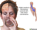
- Right cerebral hemisphere -...
- Role of the vagus nerve in ...
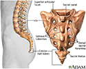
- Sacrum
- Saturated fats
- Sciatic nerve
- Sciatic nerve damage
- Scoliosis
- Scoliosis
- Scoliosis brace
- Shaken baby symptoms
- Shingles
- Signs of scoliosis
- Simple carbohydrates
- Skeletal spine
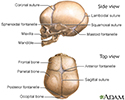
- Skull of a newborn
- Skull of an adult
- Sleep patterns in the young...
- Sleep studies
- Slit-lamp exam
- Soluble and insoluble fiber
- Spinal anatomy
- Spinal cord injury
- Spinal curves
- Spinal fusion
- Spinal stenosis
- Spinal stenosis
- Spinal tumor
- Spine supporting structures
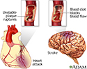
- Stroke
- Subdural hematoma
- Subdural hematoma
- Substantia nigra and Parkin...
- Sulfonylureas drug
- Systemic lupus erythematosus
- Tension-type headache
- Thiazolidinediones
- Thoracic outlet anatomy
- Tibial nerve
- Tick - deer engorged on the skin
- Tick, deer - adult female
- Torticollis (wry neck)
- Trans fatty acids
- Transient Ischemic attack (TIA)
- Tympanic membrane
- Type I diabetes
- Ulnar nerve damage
- Ultrasound, normal fetus - ...
- Untreated hypertension
- Vascular headaches
- Ventricles of the brain
- Vertebra and spinal nerves
- Vertebra, cervical (neck)
- Vertebra, lumbar (low back)
- Vertebra, thoracic (mid back)
- Vertebrae
- Vertebral column
- Vertigo
- Visual acuity test
- Visual field test
- Vitamin B6 benefit
- Vitamin B6 source
- Voiding cystourethrogram
- Weight loss
- Whiplash
- Wrist anatomy
- X-linked recessive genetic ...
- X-linked recessive genetic ...
- X-linked recessive genetic ...
- X-ray
- Yo-yo dieting
Presentations
- Carotid artery surgery - series
- Carpal tunnel repair - series
- Craniotomy - series
- CSF oligoclonal banding - ...
- Lumbar spinal surgery - series
- Meningocele repair - series
- Microdiskectomy - series
- Monitoring blood glucose - ...
- Spinal fusion - series
- Spinal surgery - cervical -...
- Stroke - series
- Two person roll - series
- Ventriculoperitoneal shunt ...

 Bookmark


